Density
In a regular photograph, the brighter parts of the scene display brighter in the print. An X-ray is a negative image. Dense objects such as bone and metal fragments display brighter because they block more of the beam reducing the film's exposure. After film development, these regions are more transparent and display brighter on the view box. In between the extremes are various shades of gray representing various sizes and densities of tissue. Objects that block X-rays are said to be radiopaque. Those that pass X-rays are radiolucent.
Unlike a photographic image, which typically registers only the object's surface features, an X-ray registers all the object's features, inside and out. Think of each point in an X-ray image as a summation of shadows. Consider all of the objects struck by a particular ray, such as bone, tissue, air pockets, and blood on its way to the film. The brightness of that point on the film is the summation of all the densities the ray encountered. A ray that passes through bone and tissue records a brighter image than a ray that passes through just bone. A ray that grazes the edge of the skull travels over a large distance of bone, which makes that part of the image very bright. A ray that traverses thin bone, such as the area around the temples on a lateral X-ray, records a darker gray image.
A normal tissue in the neck may show up as a particular shade of gray. The same tissue with air leakage into it (subcutaneous emphysema) will increase the volume of the tissue. Density is a function of mass and volume. Increasing the volume lowers the density. Thus, this tissue will show up as a darker shade of gray than the normal tissue. Radiologists refer to dark areas caused by air in the path as "air shadows."
X-Ray Reproductions – Why Books aren’t the Best Way to View X-rays
A radiologist needs to see the full range of densities, that is, shades of gray from white to black, to make a proper diagnosis. Some X-rays can have black areas that are over 500 times darker than the brightest areas, although the typical range in the tissues of interest is much smaller than this, about 32 to 1. (The ratio of black to white is called the contrast ratio.)
X-rays are transparent like photographic slides rather than prints. Radiologists read X-rays on a lighted view box. This way of displaying the image presents a much greater contrast and tonal range than a photographic print. Movies look much more vivid than photos for the same reason.
An X-ray that has been copied and published in a book will have to sacrifice many of the gray levels that are in the original X-ray. Even a good quality photograph of an X-ray will not have a contrast ratio higher than about 63 to 1. A reproduction in a book will be even lower. These ratios are significantly less the than full range of gray levels contained in the original X-rays. This means that an X-ray reproduced to show good detail in the brighter areas, such as bones, will render darker areas, such as tissues, as all black. And if the publisher adjusts the gray levels so that both the brightest and the darkest areas reproduce, then detail will be lost in the middle gray levels. One will not be able to distinguish among the fine shades of gray visible on the original X-ray. Certain features will be undetectable.
Dr. Mantik made optical density measurements of the original JFK skull X-rays at the National Archives. He found them to be unusually contrasty, with contrast ratios approaching 1000 to 1. Thus, one needs to be careful not to conclude that a large section of black in a published JFK X-ray necessarily means bone is missing from that section. To compensate for the excessive contrast, the HSCA scanned some of the X-rays into a computer and adjusted the gray levels to fit the areas of interest into a more readable range of values. These X-rays are called the enhanced X-rays.
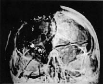
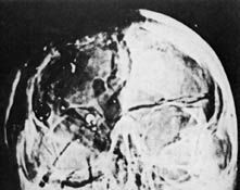
The left image is the non-enhanced AP as published by the HSCA. The middle image is the enhanced version as published in Assassination Science and the right image is the enhanced version as published in Lattimer's Kennedy & Lincoln. Notice how more of the head's upper right quadrant becomes visible in each version. This demonstrates why one needs to be careful not to infer too much from the X-rays published in books.
Projection Angle
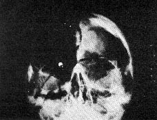
Remember that when the technicians took the AP X-ray that rigor mortis locked the head in a tilted-back position. The X-ray beam traveled through the skull in, approximately, a 30-degree upward direction (called a Modified Waters projection). This means we do not see the skull straight on in the X-ray. A point on the back of the head, such as the EOP, will be lower relative to a point on the face, such as the bridge of the nose. (The EOP region falls below the bottom edge of the enhanced AP X-ray. On the non-enhanced original it is also not visible because the dense petrous bone blocks it.)
To understand this perspective better, it is helpful to rotate the lateral X-ray to approximate the tilt of the head. The AP X-ray is on the left in the diagram below. The lateral X-ray is to its right, rotated 23 degrees counterclockwise. I drew lines to show the correspondence between various features. They won't match up exactly because the X-rays (traveling from right-to-left here) actually fan out a bit and are not parallel as drawn.
For further information, see Scaling and Orienting the X-rays
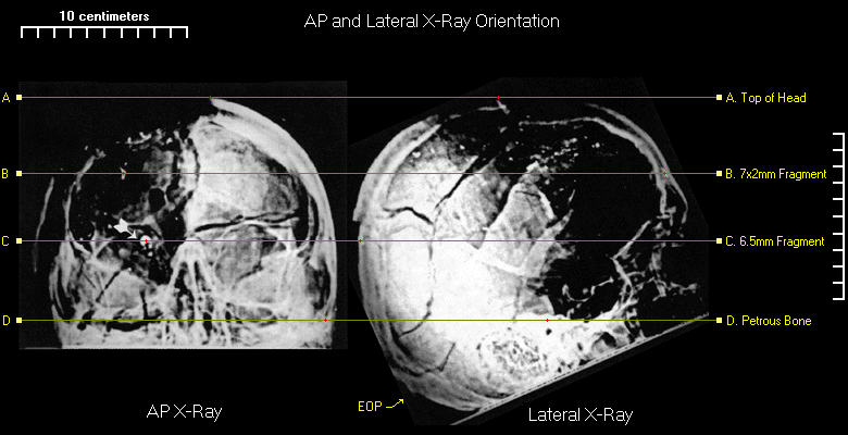
Also, some of the features show up too dark in these particular reproductions of the X-rays. The line marking the very top of the skull does not show up well here in the lateral X-ray, for example.
Clarity
Locations of the body closer to the film display greater distinctness and clarity in the image compared to those farther away. An X-ray doesn’t normally show depth, but a radiologist can use the effect to locate fractures on the near or on the distant side of the head. On the AP X-ray, the radiating fractures in back of the head, in the occipital bone, are clear with well-defined edges.
Magnification
When the X-ray tube is close to the patient, the image will be distorted because of magnification. Points on the body farther away from the film show up larger than points closer to it. The X-ray tube needs to be at least 72 inches away to minimize magnification effects. The portable unit Custer and Reed used had its X-ray tube 44 inches from the film. This means, for example, that the orbits (the eye sockets) on the AP X-ray are around 20% larger compared to a given distance on the back of the skull.
If the face is 8 inches away from the film, then the formula to calculate magnification of the facial plane is: 44" / (44" - 8") = 1.22
| Back to Taking the X-rays | Contents | Continue to Features Noted by the HSCA... |